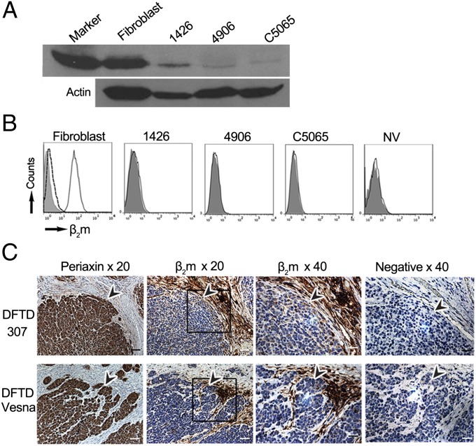
Fig. 1.
DFTD cells have low levels of intracellular MHC class I and no surface expression of β2m in vitro and in vivo. (A) Western blot of fibroblast and DFTD whole-cell protein probed with MHCI-mAb, with the MHC class I band shown at 40 kDa for a 20-min exposure to X-ray film. (A Lower) A loading control probed with an antibody to β-actin. By Bradford assay for protein, 35 µg from fibroblast lysate and 50 µg from each DFTD lysate were loaded on the gel. (B) Flow cytometry of fibroblast and DFTD cells, with fluorescence intensity on x axis and number of cells (counts) on y axis. Shaded area, stained with the preimmune serum; solid black line, stained with β2m-Ab; dashed black line, stained with β2m-Ab blocked with native devil β2m protein. (C) IHC on serial sections of primary DFTD biopsies from wild devils stained with an antibody to periaxin (a marker specific for DFTD cells, enabling DFTD cells to be distinguished from host devil cells), devil β2m-Ab, and the preimmune rat serum as a negative control for the devil β2m-Ab. Boxes indicate areas shown at 40× magnification, and arrowheads indicate similar positions in the serial sections, pointing toward DFTD cells as defined by periaxin staining. Positive cells for each marker are stained brown; nuclei are stained blue. (Scale bars: 20× magnification, 50 μm; 40× magnification, 20 μm.)

