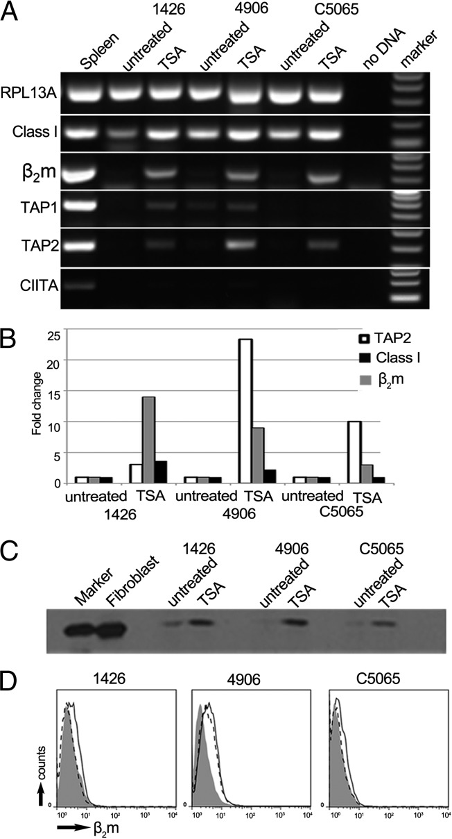
Fig. 3.
MHC class I protein is up-regulated in DFTD cells after treatment with the deacetylation inhibitor, Trichostatin A (TSA). (A) RT-PCR amplification of RPL13A, MHC class I, β2m, TAP1, TAP2, and CIITA from RNA of TSA-treated and untreated cells. Amplicons are between 100 and 300 bp. (B) RT-qPCR of TAP2 (white), β2m (gray), and MHC class I (black) in the tumor lines after treatment with TSA. Fold change is relative to the untreated control cells for each cell line and normalized against RPL13A as a housekeeping gene. A standard curve was constructed from fibroblast expression. DFTD samples were tested in triplicate. (C) Western blot of whole-cell protein from treated and untreated DFTD cells, probed with MHCI-mAb, showing MHC class I at 40 kDa. By Bradford assay for protein, 24 µg from lysate of fibroblasts, 25 µg from lysate of untreated DFTD cells, and 20 µg from lysate of treated DFTD cells were loaded on the gel. (D) Flow cytometry to test for surface expression of β2m on TSA-treated DFTD, with fluorescence intensity on x axis and number of cells (counts) on y axis. Shaded area, stained with serum from a preimmunized rat; dashed line, untreated cells stained with β2m-Ab; solid black line, TSA-treated cells stained with β2m-Ab.

