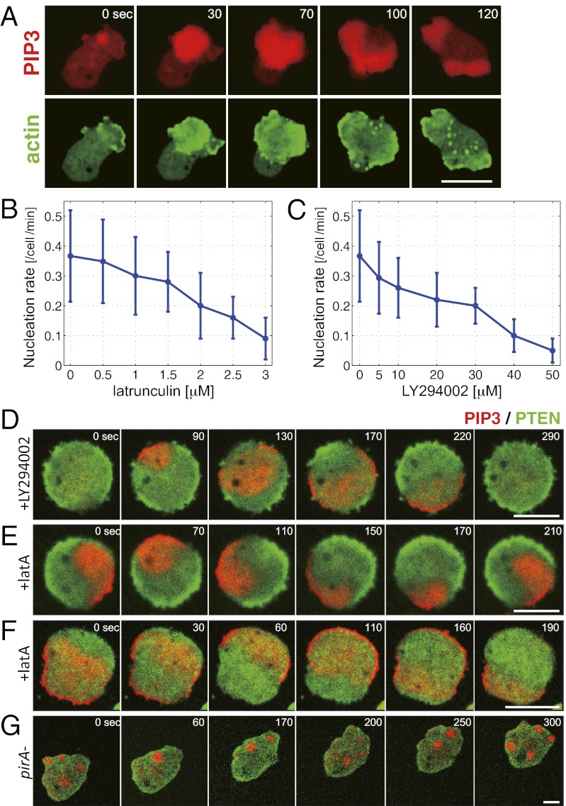
Fig. 1.
Inhibition of actin polymerization or PI3Kinase activity reduces the rate of wave nucleation and simplifies the wave patterns. (A) PIP3 (PHcrac-RFP) and F-actin (LimEΔCoil-GFP) waves at the basal membrane. (B and C) Wave nucleation rate depended on F-actin and PI3Kinase. Wave nucleation was suppressed in cells treated with latrunculin A (B) and PI3Kinase inhibitor LY294002 (C) in a dose-dependent manner. (D–G) Wave patterns in pharmacologically treated cells and a pirA− cell. (D) Transient waves were observed in cells treated with 60 μM LY294002. Rotational (E) and reflecting (F) waves were observed in cells treated with 5 μM latrunculin. (G) Sporadic localized accumulation of PIP3 in a pirA− cell. Red and green colors indicate fluorescence intensity of PH-cracRFP and PTEN-GFP, respectively. (Scale bars: 5 μm.)

