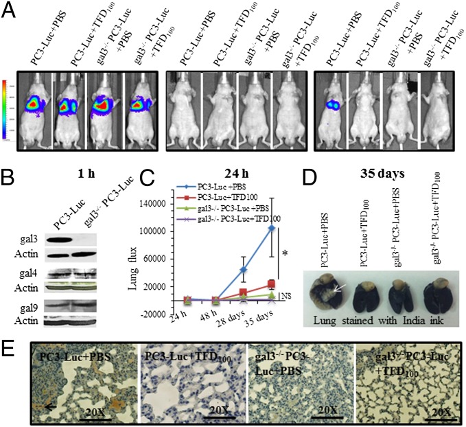
Fig. 6.
Cancer metastasis induced by PC3 cells expressing a luciferase reporter (PC3-Luc) cells and its inhibition with TFD100. (A) Representative images showing glowscale of luciferase-expressing PC3 cells (wild type or gal3 knockout) injected into the tail vein of nude mice. (B) Western blot showing expression of gal3, gal4, and gal9 in wild-type and gal3−/− (knockout) PC3-Luc cells. (C) Lung photon flux from untreated and TFD100-treated mice. NS, not significant. (D) Lungs of PBS or TFD100-treated mice stained with India ink. White arrow in the PC3-Luc+PBS injected mouse lung indicates tumor. (E) Representative diagrams showing ki67 stain of lung sections from untreated or TFD100-treated mice.

