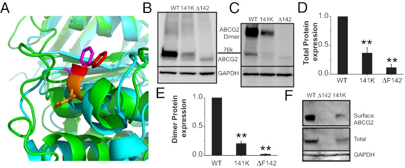
Fig. 2.
Q141K mutation occurs in mutational hotspot of ABC protein NBD. (A) Zoom-in view of superimposition of ABCG2 model (green) and human CFTR NBD1 crystal [cyan, Protein Data Bank (PDB) ID: 2PZE]. Orange, ABCG2 Q141; red, ABCG2 F142; magenta, CFTR F508. Western blots of transient total (B) and dimer expression (C) of WT, Q141K, and ΔF142 ABCG2 in HEK293 cells. Summary data of total expression of ABCG2 variants (D; n = 7 for each) and dimer expression (E; n = 6 for each). (F) Western blot of biotinylated surface and total ABCG2 expression. All means are ± SEM; **P < 0.01.

