Fig. 5.
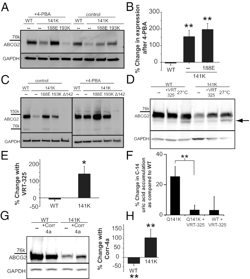
Small-molecule correction of gout-causing Q141K ABCG2 expression defect. (A) Western blot of ABCG2 variant expression in HEK293 cells demonstrating the efficacy of a 48-h treatment with 6 mM 4-PBA to correct mutant expression. (B) Summary data of 4-PBA rescue of Q141K expression (n = 3–6); 4-PBA has no effect on WT ABCG2 expression (P < 0.28; n = 4) compared with zero. (C) Western blots of ABCG2 variant dimer expression demonstrating efficacy of 48-h treatment with 6 mM 4-PBA to correct mutant dimer expression (n = 4). (D) Expression of WT and mutant ABCG2 in CHO cells and rescue with 24-h treatment with 10 µM VRT-325 or 27 °C incubation. Black arrow highlights the increase of unglycosylated protein with 27 °C incubation. (E) Summary data demonstrating efficacy of 24-h, 10-µM VRT-325 treatment for the rescue of mutant ABCG2 expression (n = 4). (F) Functional assay of ABCG2 function: percentage increase in accumulation of C-14 labeled uric acid in 2 h compared with WT accumulation (n = 5–11); higher accumulation represents lower efflux rates. (G) Western blot and summary data (H) of total Q141K ABCG2 expression rescue by 24-h treatment with 20 µM corr-4a (n = 4). GAPDH or actin expression used as a loading control on Western blots. All means are ± SEM; *P < 0.05; **P < 0.01.

