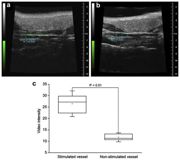
Fig. 5.
Molecular sonograms of carotid artery vessels. (a) Ultrasound imaging to detect the CD81 expression for the phenazine methosulfate (PMS)-stimulated vessels. (b) Ultrasound imaging to detect the CD81 expression for the non-stimulated vessels. Molecular imaging signal from attached ultrasound contrast agents (UCA) was color coded as green signals overlaid on gray-scale ultrasound image. There was a great enhancement in the ultrasound signal from the CD81-targeted UCA retained in the PMS-stimulated vessel wall, compared with the non-stimulated vessel wall. (c) Box plot of video intensity for the stimulated and non-stimulated carotid artery vessels. The selected contrast regions were marked with boxes.

