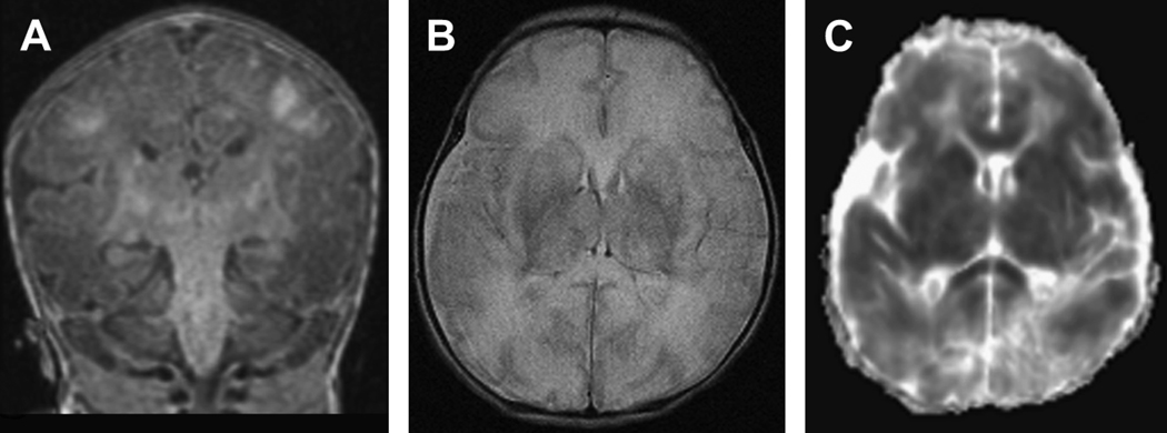
Figure 1.
Diffuse neuronal injury in a term infant with a history of severe hypoxic-ischemic injury. Images were acquired at 7 days of life. A) Coronal T1-weighted image shows interruption of the posterior limbs bilaterally with diffuse hyperintesities in the watershed regions of the gyri in a para-sagittal distribution. In addition, one can visualize the high signal in the deep nuclear gray matter. B) Axial T2-weighted image shows injury throughout the deep nuclear gray matter with diffuse white matter hyper intensity and loss of differentiation between white matter and the cortical ribbon. C) Axial diffusion-weighted image where restricted diffusion from injury (decreased ADC) is represented by dark lesions. This image shows diffuse diffusion restriction in the deep nuclear gray matter, white matter, and cortex bilaterally still apparent at day 7.

