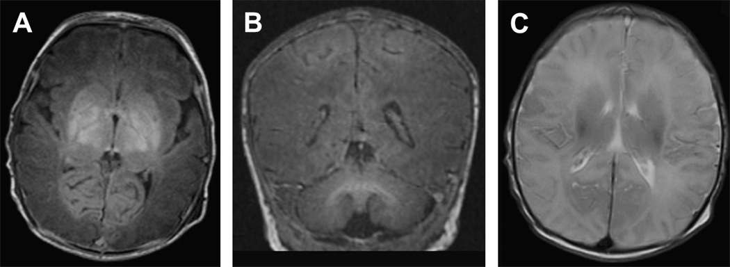
Figure 2.
Deep nuclear gray matter with cortical injury in a term infant with a history of severe hypoxic-ischemic injury. A) Axial T1-weighted image shows hyper intense severe injury throughout the deep nuclear gray matter. B) Coronal T1-weighted image shows the extension of this hyper intense injury from the deep nuclear gray matter up into the parasagittal region of the cortex. C) Axial T2-weighted image demonstrates low intensity extending from the DNGM into the parasagittal cortex bilaterally.

