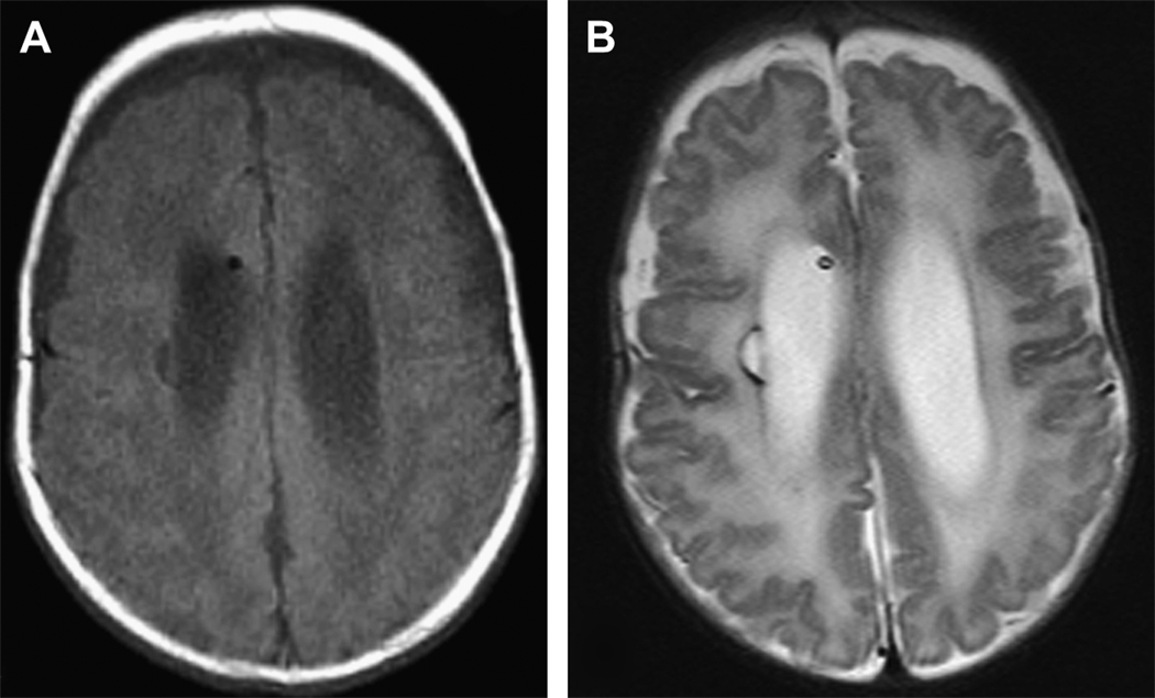
Figure 3.
Periventricular leukomalacia in a former 26 week male with grade III IVH and post hemorrhagic hydrocephalus requiring a shunt. Images were acquired at term equivalent. A) Axial T1-weighted image shows a focal cystic hypo intensity along the right periventricular region. B) Axial T2-weighted image shows this injury as a hyper intense lesion again along the right periventricular area. Furthermore, there is diffuse hyper intensity of the white matter bilaterally consistent with diffuse as well as focal periventricular leukomalacia.

