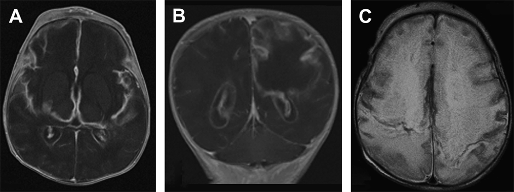
Figure 6.
Severe multi-cystic encephalopathy in a 12 day old term infant with proteus meningitis. The first group (A,B) are T1-weighted axial (A) and coronal (B) images showing severe white and gray matter loss in the frontal, parietal, and temporal lobes. This tissue loss is replaced largely by fluid. (C) T2-weighted axial image also shows diffuse injury with necrosis of both white and gray matter.

