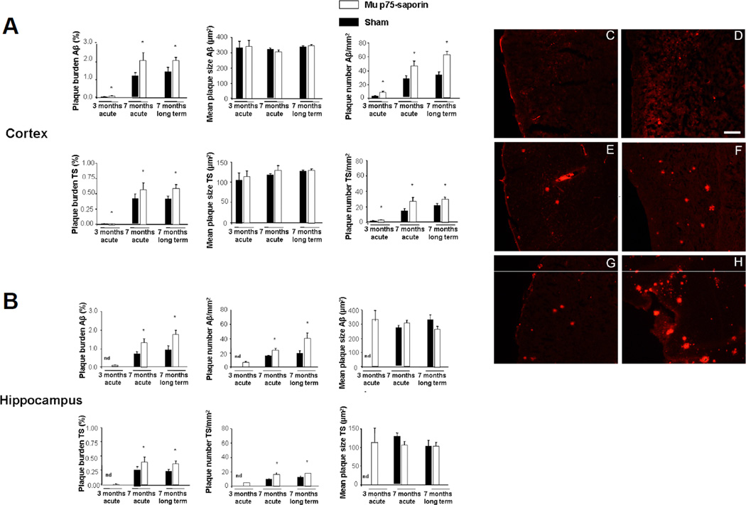
Figure 5.
Postmortem assessment of senile plaque (SP) deposition with anti-β-amyloid (Aβ) antibody and thioflavin S (TS) staining in cortex and hippocampus of APP/PS1 mice with or without murine p75NTR saporin (mu p75-SAP)-induced lesions. (A, B) There was a significant increase in plaque burden in all lesioned mice, although this effect was greater in 7-month-old mice, particularly in those with long-term lesions. The increase in amyloid burden, both in cortex (A) and hippocampus (B) was not due to increased plaque size but to an increase in the number of new deposited SPs. Data are representative of 3 to 6 mice per group. Statistical differences were detected by Student t-test for independent samples and Mann-Whitney U-test for independent samples in the case of number of plaques per mm2. * p < 0.05. (C–H) Examples of Aβ immunostaining in the cortex: C, Sham 3 months acute; D, Mu p75-SAP, 3 months acute; E, Sham 7 months acute; F, Mu p75-SAP, 7 months acute; G, Sham 7 months long-term; H, Mu p75-SAP 7 months long-term. Scale bar = 250 µm.

