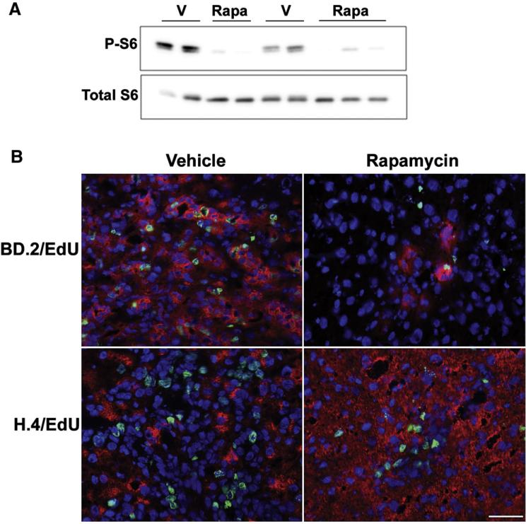
Fig. 6.
Effect of rapamycin treatment on oval cell abundance and proliferation in rats treated with 2-AAF followed by 2/3 partial hepatectomy (PHx). (A) Western immunoblotting for phospho-S6Ser235/236 and total S6 was performed on total liver homogenates from vehicle (V) or rapamycin (Rapa) treated rats placed on the 2-AAF PHx protocol and harvested 7 days post-hepatectomy. (B) Cryosections from these animals underwent immunofluorescent staining for an oval cell marker BD.2 and a marker of DNA synthesis EdU or (C) a hepatocyte marker H.4 and EdU. Slides were counterstained with DAPI. Images were acquired at 40×. Scale bar: 50 μm. Image analysis was undertaken as described in the Supplemental Information.

