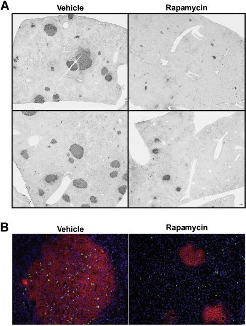
Fig. 7.
Effect of rapamycin treatment on the abundance and size of preneoplastic foci in rats placed on the Solt–Farber protocol. Rats subjected to the Solt–Farber protocol were treated with rapamycin or DMSO vehicle and liver harvested 7 days post-hepatectomy. (A) Sections from these animals were stained with anti-GST-P. Images from duplicate animals were acquired at 2×. Scale bar: 50 μm. (B) Cryosections from these animals underwent immunofluorescent staining for GST-P and a marker of DNA synthesis EdU. Slides were counterstained with DAPI. Images were acquired at 10×. Scale bar: 50 μm. Image analysis was undertaken as described in the Supplemental Information.

