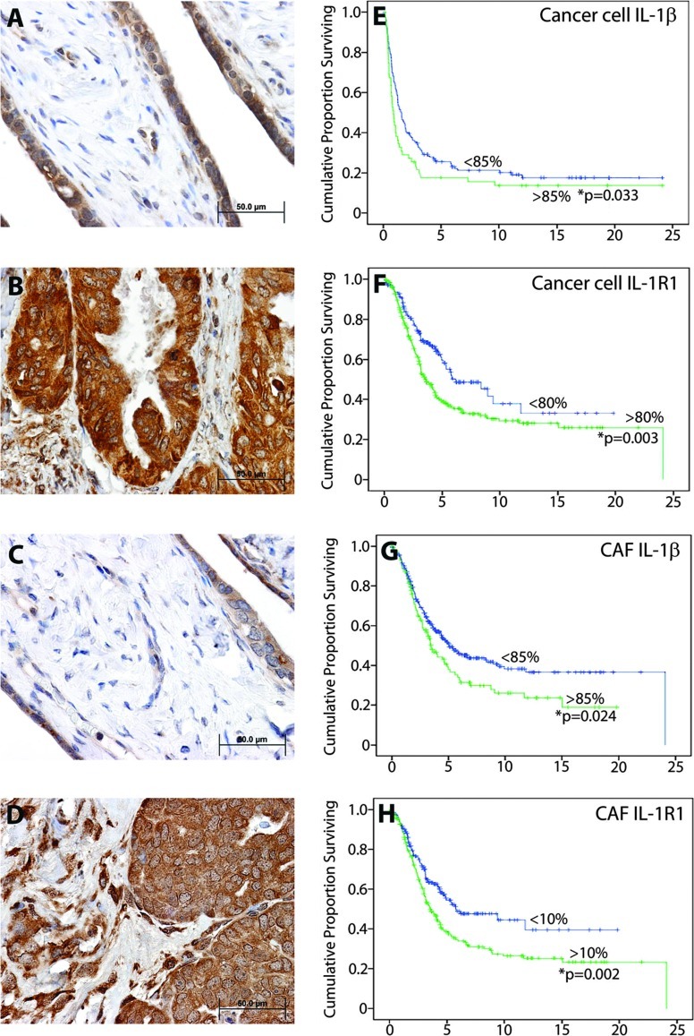
Figure 2.
The IL-1β/IL-1R1 signaling axis is highly elevated in ovarian cancer and correlates with poor ovarian cancer patient survival. Background IL-1β IHC detection is observed in both the epithelium and stromal compartment of normal ovarian cyst (A), as opposed to highly elevated IHC staining in both compartments in ovarian cancer tissue (B). IL-1β cognate receptor IL-1R1 displays a similar pattern of background detection in normal ovarian cyst tissue (C), compared to elevated staining in ovarian cancer epithelial cells and CAFs (D). Kaplan-Meier patient overall survival analysis curves (E–H) for ovarian carcinoma patients show reduced survival time as a function of (E) ≥85% IL-1β expression in epithelial cancer cells (P = .033; N = 397), (F) ≥80% IL-1R1 expression in epithelial cancer cells (P = .003; N = 406), (G) ≥85% IL-1β expression in ovarian cancer fibroblasts (P = .024; N = 204), and (H) ≥10% IL-1R1 expression in ovarian cancer fibroblasts (P = .002; N = 231).

