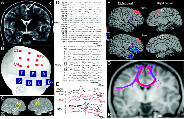
Fig 1.
A, MR image shows a right superior frontal gyrus lesion (arrow). B, Active sites that show frequent spikes on EEG and MEG. Red circles and blue squares show EEG electrodes and MEG sensors, respectively. Note that the active MEG electrodes are located more inferiorly than the active EEG electrodes. C, The location of grids is shown on the cortical surface. D, A typical spike recorded on simultaneous EEG and MEG. The labels of the MEG channels correspond to the sensors shown on B, Each site has 2 gradiometers. E, Source waveforms of an EEG/MEG spike and a typical spike on IEEG. Source waveforms are extracted at sites 1 and 2 (superior frontal) for EEG and 3 and 4 (inferior frontal) for MEG. The superior frontal peak of EEG waveforms precedes the inferior frontal peak of MEG waveforms by approximately 20 ms. A similar time difference is seen on the IEEG spike. F, A source distribution map of a typical spike appearing on both EEG and MEG. Cortical activation is shown with red/yellow for EEG and blue/dark blue for MEG. The EEG activation appears in the right superior frontal area at the early latency (0 ms), whereas the MEG source shows later activation in the inferior frontal area (14 ms). White and red circles show the location of active IEEG electrodes in the superior (sites 1 and 2) and inferior (sites 3 and 4) frontal areas, respectively. G, The tractography shows a fiber connection between the right superior and inferior frontal gyri.

