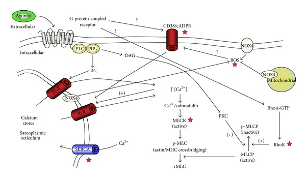
Figure 2.

Overview of the signalling pathways involved in airway smooth muscle contraction. The contractile agonist interacts with its specific G-protein-coupled receptor (GPCR) leading to the activation of phospholipase C (PLC) which hydrolyzes phosphatidylinositol bisphosphate (PIP2) leading to the formation of two-second messengers, inositol triphosphate (IP3), and diacylglycerol (DAG). IP3 interacts with its receptor (IP3R) on the sarcoplasmic reticulum (SR) leading to the release of calcium ions Ca2+ which in turn, through forming a complex with calmodulin, activates myosin light chain kinase (MLCK). MLCK phosphorylates regulatory myosin light chain (rMLC) to form p-MLC which leads to myosin and actin crossbridging and contraction. p-MLC is deactivated by the action of myosin light chain phosphatase (MLCP). Both DAG, through its action on protein kinase C (PKC), and RhoA, through its target Rho Kinase (RhoK), have an inhibitory action on MLCP through phosphorylation. Interaction of agonist with GPCR also activates both CD38/cADPR and Rho/RhoK pathways, although the exact mechanism is not fully known. CD38/cADPR activation leads to the release of Ca2+ from SR through ryanodine receptors (RyR) channels. Nicotinamide adenine dinucleotide phosphate oxidase type 4 (NOX4) generates reactive oxygen species (ROS) which may affect ASM calcium homeostasis and subsequently ASM contraction through their action on the CD38/cADPR pathway. Red stars ★ indicate signalling points with abnormalities suspected of driving hypercontractility in asthmatic ASM.
