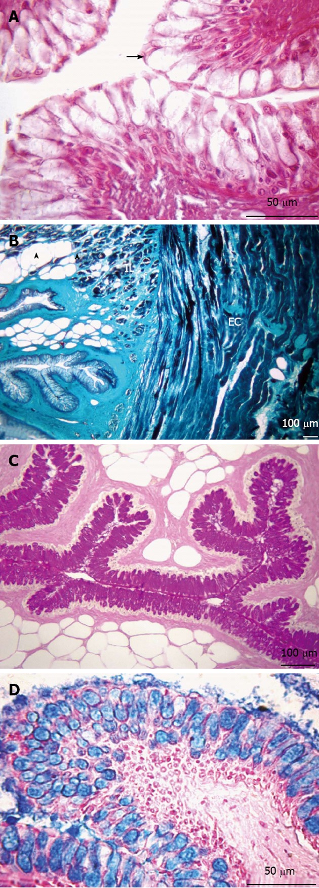
Figure 2.

Transversal sections of the esophagus. A: Non-keratinized stratified squamous epithelium (arrow). Hematoxylin and eosin stain; B: Presence of adipose tissue (arrowheads) in the submucosa layer. Muscular layer formed by two sub-layers, internal longitudinal (IL) and external circular (EC). Gomori’s trichrome stain; C: Presence of neutral glycoconjugates (GCs). Periodic acid-Schiff stain; D: Acid GCs. Alcian blue stain.
