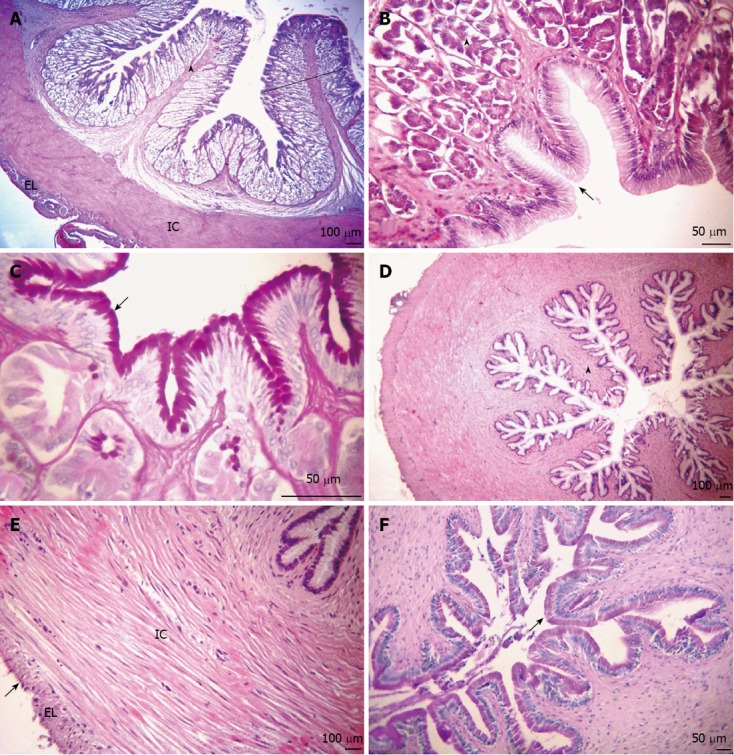
Figure 3.

Transversal sections of the stomach. A: Mucosa layer with many high gastric folds (arrowhead), thickening of the lamina propria caused by a large number of tubular gastric glands (outline). Organization of the internal circular (IC) and external longitudinal (EL) muscular layers. Hematoxylin and eosin (HE) stain; B: The simple columnar epithelium with mucus-secreting cells forming faveola gastrica (arrow) and fundic glands composed of oxynticopeptic cells (arrowhead). HE stain; C: Presence of neutral glycoconjugates (GCs) (arrow). Periodic acid Schiff (PAS) stain; D: Mucosa layer, together with the sub-mucosa and internal muscular layers, forming large longitudinal folds (arrowhead). HE stain; E: The muscular layer of smooth muscle fibers, composed of IC and EL sub-layers and serosa (arrow). HE stain; F: Neutral GCs (arrow). PAS stain. (A-C: Glandular region; D-F: Non-glandular region).
