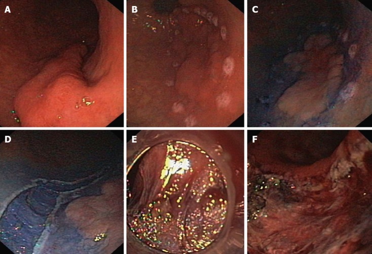
Figure 1.

Endoscopic submucosal dissection of gastric neoplastic lesion. A: Lesion type IIa+c localized in the antrum; B: Margins of the lesion border were marked with the needle knife; C: Solution of indigo carmine in saline was injected into the submucosal space; D: An incision was made in the mucosa and submucosa around the lesion with normal mucosal margin; E: Submucosal dissection performed directly under vision control; F: Mucosal defect after the completed procedure.
