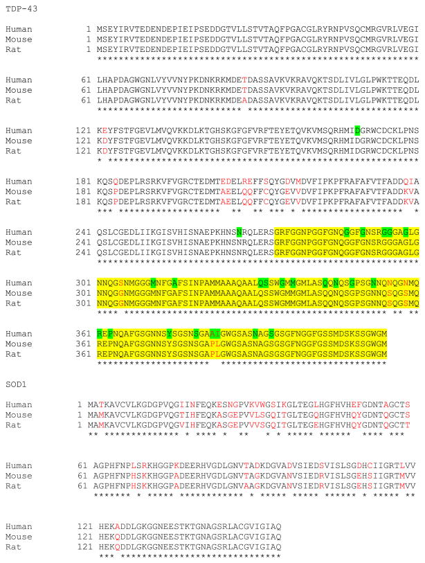
Fig. 1.
Multiple sequence alignments for TDP-43 and SOD1. Amino acid sequences are compared for TDP-43 (RefSeq ID for human: NP_031401.1; mouse: RefSeq ID: NP_663531.1; GenBank no. for rat: EDL81132.1) and SOD1 (RefSeq ID for human: NP_000445.1; mouse: NP_035564.1; rat: NP_058746.1). Nonidentical residues are shown in red text. The yellow highlighted area marks the TDP-43C-terminal domain, and the green highlighted residues mark the positions of ALS-associated mutations in human TDP-43.

