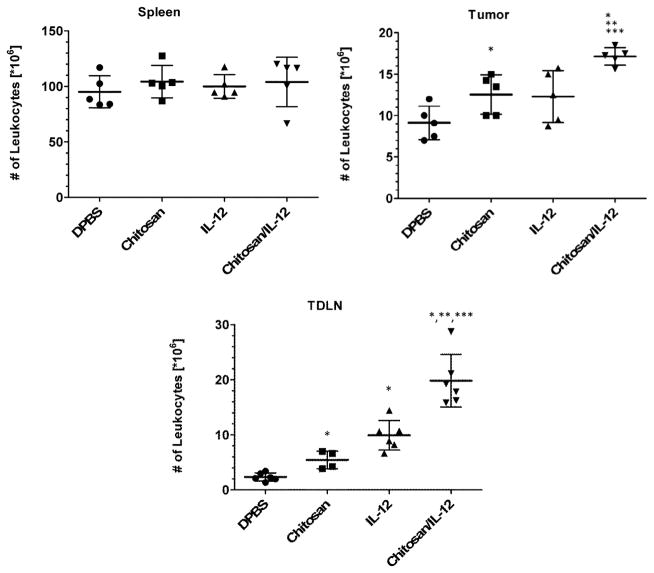
Fig. 3.
Effect of treatment on leukocyte expansion in different tissues. C57BL/6J mice (n=6 per group) bearing MC38 flank tumors received i.t. injections of DPBS, chitosan alone, IL-12(1μg) alone or chitosan/IL-12(1μg) on days 7 and 14. On day 17, spleens, tumors and tumor draining lymph nodes (TDLNs) were harvested and viable leukocytes enumerated via automated cell counting. Data for individual mice as well as mean ± standard deviation are presented. *P<0.05 vs. DPBS; ** P<0.05 vs. chitosan; *** P<0.05 vs. IL-12.

