Fig. 3.
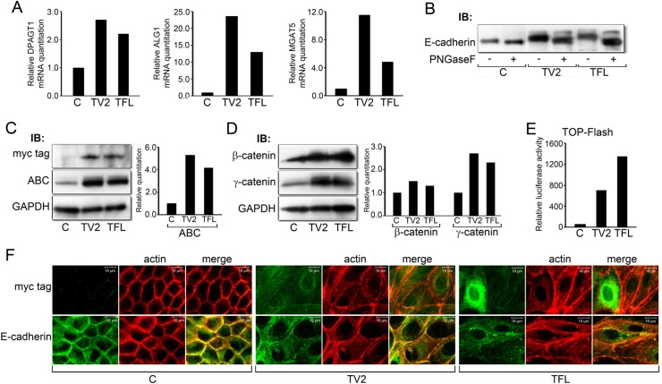
Expression of recombinant DPAGT1 drives canonical Wnt signaling and N-glycosylation of E-cadherin. (A) Quantitative PCR of DPAGT1, ALG1 and MGAT5 transcripts from cells transfected with the full length (TFL) and variant 2 (TV2) DPAGT1 cDNA clones. Results represent one of two independent determinations. (B) N-glycosylation of E-cadherin in control cells (C) and TV2 and TFL transfectants before and after PNGaseF treatment. Results are representative of three independent experiments. (C) Immunoblot of recombinant GPT isoforms (Myc tag) and ABC from control cells (C) and TV2 and TFL transfectants. Bar graph: fold change in ABC levels after normalization to GAPDH. Results are representative of one of two independent experiments. (D) Immunoblot of β- and γ-catenins from control cells (C) and TV2 and TFL transfectants. Bar graph: fold change in β- and γ-catenin levels after normalization to GAPDH. Results are representative of one of two experiments. (E) Luciferase reporter activity from the TOP-Flash vector in control (C) cells and TV2 and TFL transfectants. Results are representative of one of three independent determinations. (F) Immunofluorescence localization of GPT (Myc tag) and E-cadherin in control cells (C) and TV2 and TFL transfectants counterstained for F-actin. Scale bars, 10 µm.

