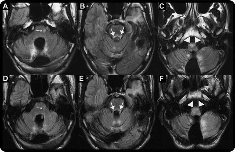
Figure. Serial brain MRI at oculopalatal tremor (OPT) onset (A–C) and following resolution of OPT (D–F).
(A, B) Axial fluid-attenuated inversion recovery (FLAIR) study shows new hyperintense signal in the pons (small arrow) and bilateral superior cerebellar peduncles (large arrows). (C) Axial FLAIR study shows hyperintense signal in enlarged inferior olivary nuclei (right greater than left) (arrowhead) consistent with hypertrophic degeneration. (D, E) Axial FLAIR study shows resolving hyperintense signal in the pons (small arrow) and bilateral superior cerebellar peduncles (large arrows). (F) Axial FLAIR study shows persistent hyperintense signal in enlarged inferior olivary nuclei (right greater than left) (arrowhead).

