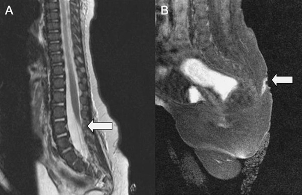
Figure 2. Spine MRI.

(A) Sagittal T2-weighted MRI of the spine shows the termination of the conus medullaris at the upper level of L4, defining a tethered cord, without an identified lipoma. (B) An inflamed lumbosacral sinus dermal tract was demonstrated with diffuse gadolinium enhancement along its course.
