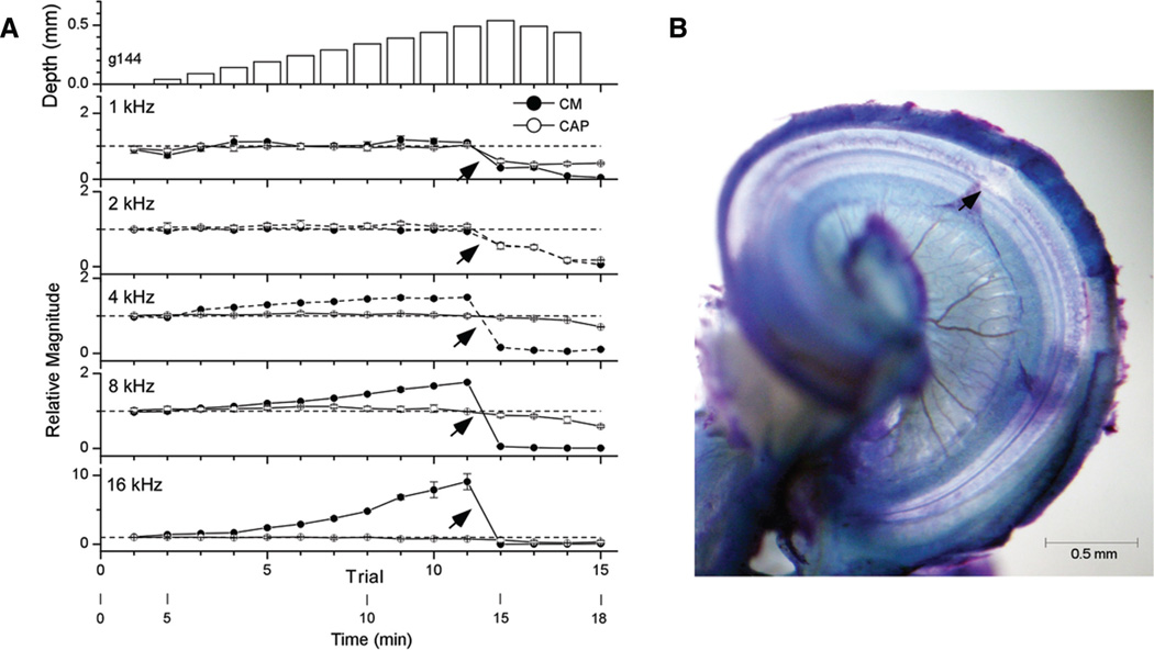
Fig. 4.
Example of an insertion of a rigid electrode with histological damage in the BM. A, Physiology. Format as in Figure 2. At trial 12 (arrows, 0.54 mm) there was a sharp decline at all frequencies. B, Histology. The arrow depicts the site of damage, which was a hole in the BM. BM, basilar membrane.

