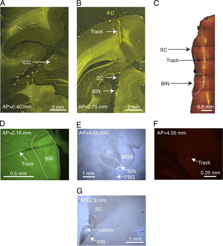
Figure 1.

A, A brain section stained for cytochrome oxidase from marmoset 12W. The ICC is present in this section, shown by the dark staining indicated by the arrow, but the BIN is not, and there are no tracks. B, Another brain section in the same animal located 2.35 mm anterior to the section in A. The arrows point to the track mark through cortex, the SC, and the BIN. C, A higher magnification of the section shown in B viewed with fluorescence microscopy. The picture is a photomontage of several adjacent images. The arrows point to the SC, the BIN (outlined), and the fluorescently labeled electrode track. D, Labeled track through the posterior right BIN in a different marmoset (18W) using a lateral electrode approach. E, A far anterior section of the right BIN from 18W also showing the SC, medial geniculate body (MGB), and the parabigeminal nucleus (PBG). The track is not labeled at this depth. F, The same section as E at a position dorsal and lateral that contains a piece of the fluorescently labeled track (arrow). G, A posterior section through the left BIN from animal 18W where an electrolytic lesion was made (arrow).
