
Figure 10.
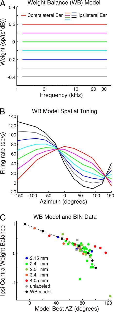
A, Contralateral weights are fixed, whereas the ipsilateral weights vary according to color for the different models. B, Firing rate versus AZ for the different WB models in A driven with VS stimuli. C, Ipsilateral–contralateral weight balance (Wic, defined in Materials and Methods) versus best model AZ. Broken lines and black dots are from the WB models in B, Colored points are for BIN data measured in different craniotomies, identified in the legend. Positions range from posterior (blue) to anterior (red). Four outliers were removed from C and the correlation analysis: two neurons had small noisy weights, and two were tuned to far posterior locations.