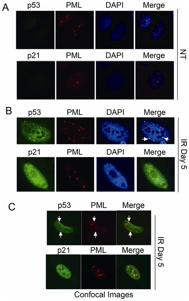
Figure 3. p53 colocalized with PML in irradiated U2OS cells.

U2OS cells on glass coverslips were either untreated (NT, A) or treated with irradiation (IR, 10Gy, B,C). Cells were fixed with 4% formaldehyde at indicated time points. P53, p21 antibodies were detected using an Alexa-488 conjugated goat anti rabbit IgG. PML antibody were detected using a rhodamine-x conjugated goat anti mouse IgG. Cell nuclei were counterstained with DAPI (blue). Images were captured at 100× magnification. Representative images were shown. Representative foci with p53/PML colocalization were pointed with white arrows. Representative confocal images were shown in C.
