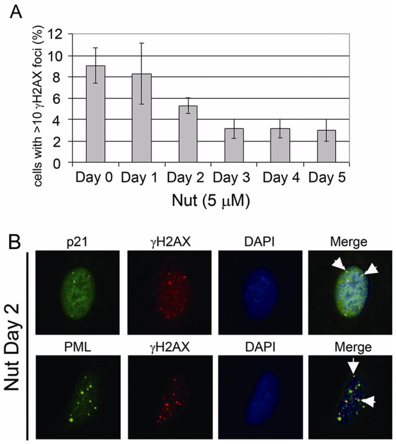
Figure 7. p21 colocalized with γH2AX in Nutlin treated U2OS cells.

U2OS cells on glass coverslips were treated as described in Figure 4. A) Percentage of cells with >10 γH2AX foci was determined at indicated time points after treatment. Percentage represents Mean ± SEM of three independent experiments with 200 cells counted per experiment. B) p21, PML antibodies were detected using an Alexa-488 conjugated goat anti rabbit IgG. γH2AX antibody were detected using a rhodamine-x conjugated goat anti mouse IgG. Representative colocalization foci were pointed with white arrows.
