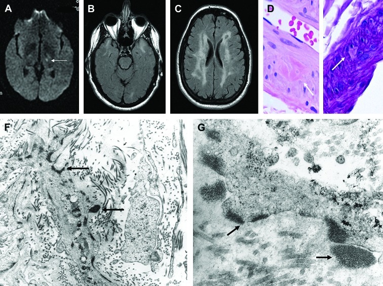
Figure 1.
Radiologic findings in patient with CADASIL disease. (A) Magnetic resonance imaging (at age 43 years, when patient developed acute-onset right hemiparesis and dysarthria), revealed left internal capsule lacunar infarct (arrow). On T2-weighted images, there were symmetric hyperintense signals in white matter of both temporal lobes (B), and diffuse hyperintense signals in subcortical and deep white matter (C). Light microscopic examination of skin biopsy specimen fixed in formalin revealed rare dermal arteriole exhibiting focal aggregates of eosinophilic material (D), which was PAS positive diastase resistant, and focally replaced smooth muscle cells of media (E). (F) and (G) On transmission electron microscopic examination of tissue fixed in glutaraldehyde, GOM deposits (black deposits marked by arrows) were identified in extracellular matrix, adjacent to and within smooth muscle cells of dermal arterioles. Some of the extracellular deposits appeared to indent cell membranes of atrophic/degenerated smooth muscle cells of media. (F and G, Original magnifications: F, x 23,000; G, x 73,000.) Reproduced with permission from Elsevier.

