
Figure 4.
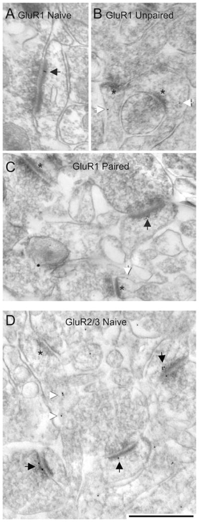
Representative electron micrographs from the dorsal LA of rats that received no stimuli (i.e., naïve), paired, and unpaired presentation of stimuli.A–C: Postembedding gold (PEG) labeling for the GluR1 subunit of AMPARs. D: PEG labeling for the GluR2/3 subunits of AMPARs. Black arrows across all panels indicate synaptic PEG labeling; white arrows indicate nonsynaptic labeling within spine heads (B,C) and spine neck (D). Asterisks point to PSDs of unlabeled synapses. Scale bar = 500 nm in D (applies to A–D).