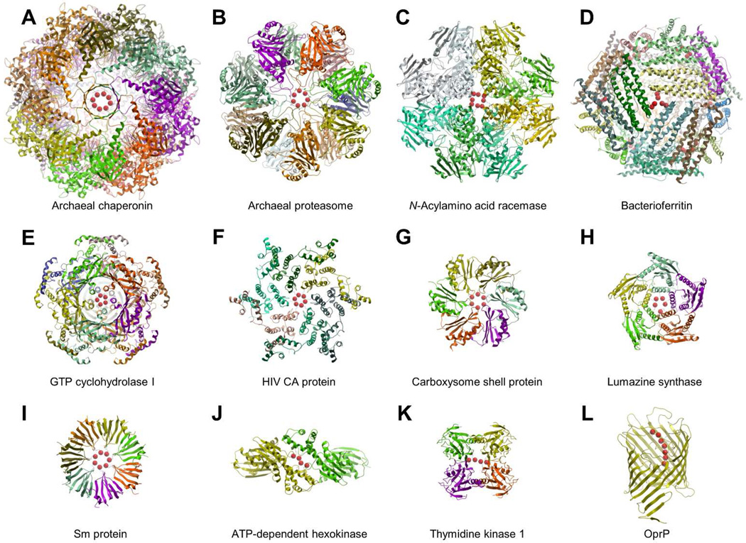
Figure 1.
Representative examples of arginine clusters with Cζ-Cζ distances < 5 Å obtained from the PDB database. A) Archaeal chaperonin (PDB entry 3kfb); B) Archaeal proteasome (PDB entry 1j2q); C) N-Acylamino acid racemase (PDB entry 1r0m); D) Bacterioferritin (PDB entry 1nf4); E) GTP cyclohydrolase I (PDB entry 1wpl); F) HIV CA protein (PDB entry 3h4e); G) Carboxysome shell protein, (PDB entry 2a10); H) Lumazine synthase (PDB entry 2f59); I) Sm protein (PDB entry 1i8f); J) ATP-dependent hexokinase (PDB entry 2e2q); K) Thymidine kinase 1 (PDB entry 2wvj); L) Outer membrane protein (PDB entry 2o4v). The protein structures are represented with a ribbon model and colored by subunit. Cζ atoms are represented in red with space filling spheres.

