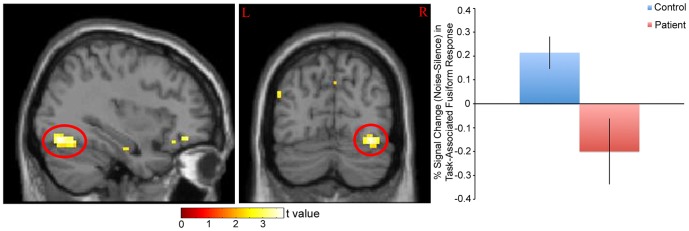
Figure 4. % Signal Change (Noise-Silence) in Task-Associated Right Fusiform Response in Patients and Controls.

Left: Statistical parametric map. Map was thresholded at p<0.01 and overlaid onto the SPM8 canonical single subject T1 image for visualization. Data are shown in the neurologic convention (R on R). Right: Extracted right fusiform response, based on the cluster circled in red on the parametric map (peak coordinate: x = 39, y = −76, z = −14). A relative increase in task-associated response in noise (compared to silence) in controls and a decrease in response in patients was observed.
