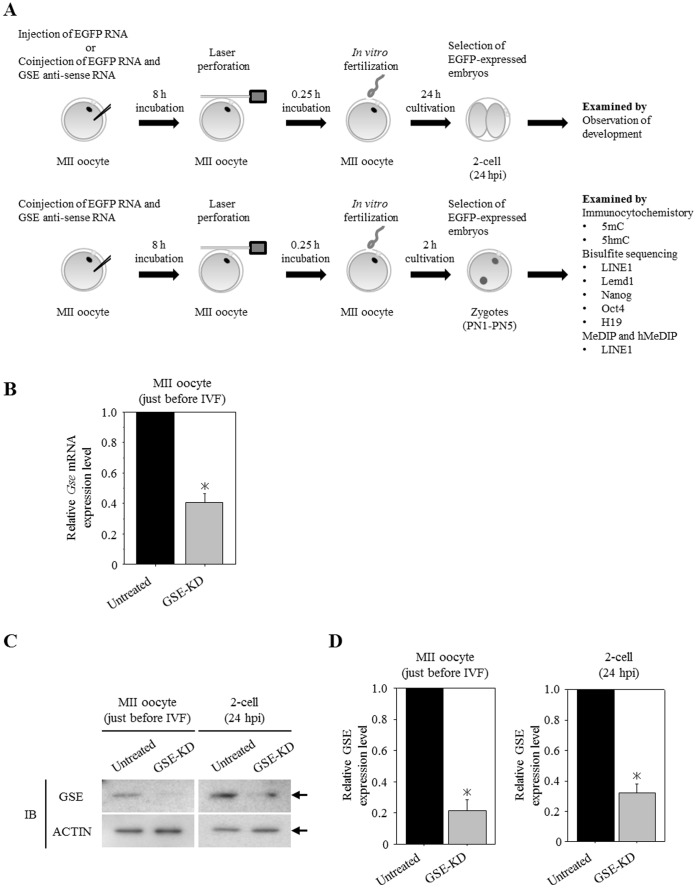
Figure 3. Maternal GSE-knockdown (GSE-KD) zygotes obtained from antisense RNA injection and in vitro fertilization (IVF).
(A) Scheme of the experimental procedures. In immunocytochemistory, bisulphite sequencing, and MeDIP and hMeDIP (methylated and hydroxymethylated DNA immunoprecipitation) analyses, EGFP RNA-uninjected MII oocytes, incubated under similar conditions, were used as untreated controls. (B) Knockdown of GSE expression by an antisense RNA was confirmed by quantitative RT-PCR analysis. The relative ratios were obtained by dividing the expression level of the Gse gene by the expression level of the G3PDH gene. More than 90 oocytes from three independent experiments were analyzed. Shown are statistically significant differences between untreated and GSE-KD oocytes (*p<0.05). Bars represent the standard error of the mean. (C) Knockdown of GSE protein expression was confirmed by immunoblot analysis of MII oocytes just before IVF or in 2-cell embryos. Ninety oocytes from three independent experiments were analyzed. Actin was used as a loading control. (D) Densitometric quantification of the immunoblot bands of Figure 3C showing statistically significant differences between untreated and GSE-KD oocytes or embryos (*p<0.05). Bars represent the standard error of the mean.

