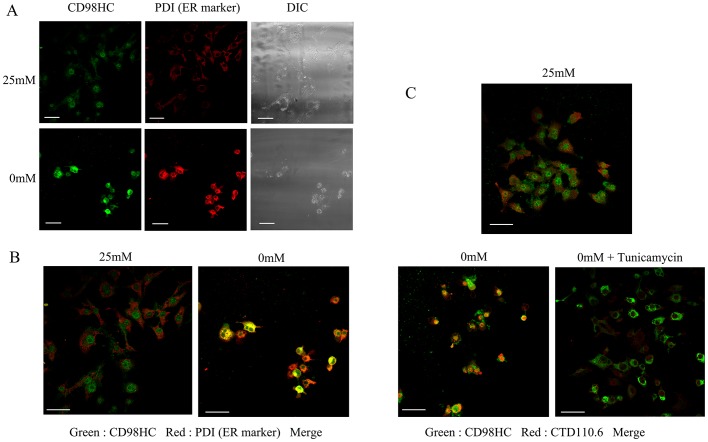
Figure 3. Subcellular localization of CD98HC protein.
A, T24 cells were cultured in either 25 mM or 0 mM glucose medium for 1day, fixed, and stained with both rabbit polyclonal antibodies for CD98HC and a mouse monoclonal antibody for PDI. The cells were then treated with Alexa 488-labeled anti-rabbit IgG and Cy3-labeled anti-mouse IgG. Fluorescence was observed by confocal fluorescent microscopy. DIC shows photographs obtained using a differential interference contrast microscope. B, Enlarged merged photographs. Note that CD98HC and PDI were co-localized under glucose deprivation. c, The effects of tunicamycin treatment on the subcellular localization of proteins that reacted with CTD110.6 and anti-CD98HC antibodies under glucose deprivation. T24 cells were cultured either in 25 mM glucose medium, 0 mM glucose-deprived medium, or 0 mM glucose with tunicamycin. The cells were fixed and stained with rabbit polyclonal antibodies for CD98HC and a mouse IgM monoclonal antibody CTD110.6. The cells were then treated with Alexa 488-labeled anti-rabbit and Cy3-labeled anti-mouse antibodies. Merged photographs are shown. Note that CD98HC and CTD110.6 were co-localized under glucose deprivation, but not under glucose deprivation in the presence of tunicamycin. The white scale bars correspond to 50 µm.

