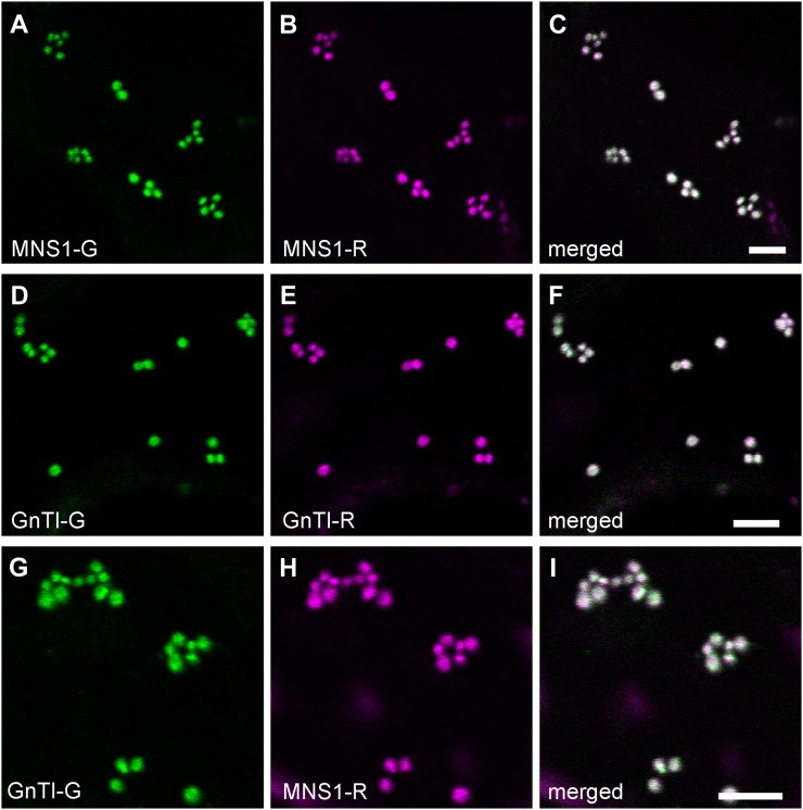
Figure 2.
Golgi localization of full-length cis/medial-Golgi protein pairs in tobacco leaves. Confocal images show representative tobacco leaf epidermal cells coexpressing fluorescent protein-tagged full-length protein pairs MNS1-G (A) and MNS1-R (B), GnTI-G (D) and GnTI-R (E), and GnTI-G (G) and MNS1-R (H). C, F, and I show merges of green (GFP fluorescence) and magenta (mRFP fluorescence) channels. White in the merged images indicates areas of colocalization. Bars = 5 µm.

