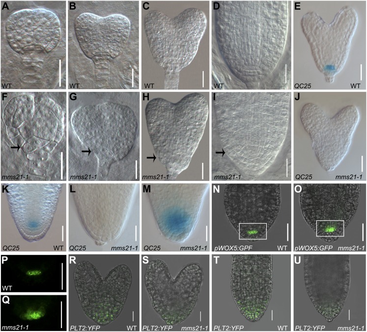
Figure 4.
mms21-1 shows defects in the root stem cell niche during embryogenesis. A to D, Wild-type (WT) embryos in late globular (A), early heart (B), middle heart (C), and cotyledon (D) stages. F to I, mms21-1 mutant embryos at stages equivalent to A to D. Arrows point to aberrant cell divisions in the basal embryo domain of mms21-1 mutants. E to J, QC25 expression in heart-stage embryos of the wild type and mms21-1. K to M, QC25 expression in mature-stage embryos of the wild type and mms21-1. N to Q, pWOX5:GFP expression in mature-stage embryos of the wild type and mms21-1 (P shows the outlined area in N and Q shows the outlined area in O, observed in the GFP channel). R to U, Expression patterns of PLT2pro:PLT2:YFP in wild-type and mms21-1 embryos. Bars = 20 μm.

