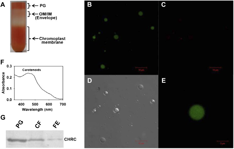
Figure 6.
Confocal microscopy imaging and western blotting of isolated plastoglobules. A, Suc density gradient purification of plastoglobules (PG). Note that the distinct upper layer of purified plastoglobules is well separated from the underlying chromoplast membranous fractions. OM/IM indicate outer and inner membranes of chromoplasts. B to D, Purified plastoglobules were visualized using a confocal microscope. On excitation with a 488-nm laser, the plastoglobules exhibit strong green fluorescence (emission between 500 and 510 nm; B) but show negligible red fluorescence (emission between 740 and 750 nm; C). D shows a bright-field image of the plastoglobules. E, Enlarged view of a single plastoglobule. Note that the green fluorescence emitted from plastoglobules is uniformly distributed. F, Absorption spectrum of the extract obtained from purified plastoglobules showing characteristic carotenoid peaks at 440 to 475 nm. G, Western analysis of CHRC levels in the purified plastoglobule fraction (PG; 0.33 μg) in comparison with the chromoplast membrane fraction (CF [the chromoplast fraction prior to sonication]; 2.5 μg) and total protein from the outer pericarp of RR fruits of hp1 (FE, fruit extract; 2.5 μg). Note that the plastoglobule fraction shows enrichment of CHRC protein. [See online article for color version of this figure.]

