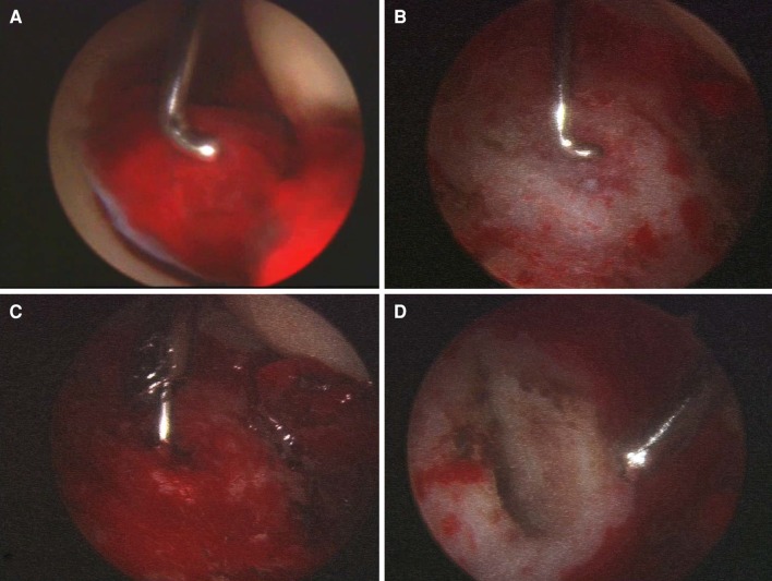
Fig. 3A–D.
These intraarticular views show (A) severe synovial proliferation around the ligamentum teres; (B) the center of the lesion, indicated by the probe after the synovectomy and débridement of the ligamentum teres were performed; (C) the lesion being heated to 90°C for 7 minutes using a rigid radiofrequency electrode with a diameter of 1 mm; and (D) the lesion after ablation of the nidus.

