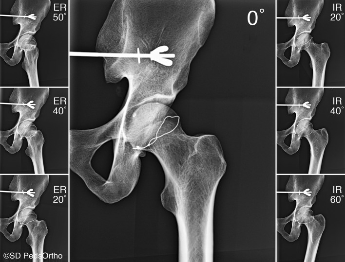
Fig. 2.
AP radiographs of the cadaver proximal femur in 50°, 40°, and 20° external rotation (ER), 0° neutral/anatomic rotation, and 20°, 40°, and 60° internal rotation (IR) are shown. A radiopaque wire had been glued to the cadaveric proximal femur to represent a theoretical growth plate for a previous unpublished study but was not used in this study.

