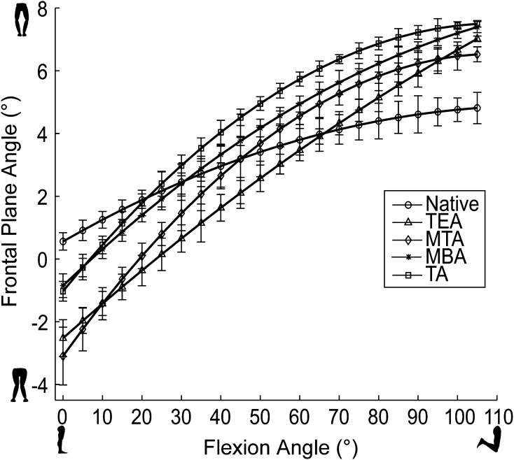
Fig. 6.
Varus-valgus kinematics during passive flexion for a representative specimen is shown. In early flexion, all alignments show more valgus motion than the native condition, but the TA and MBA alignments minimize this. TEA = transepicondylar axis; MTA = medial third axis; MBA = medial border axis; TA = transverse axis.

