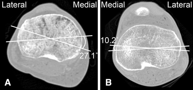
Fig. 7A–B.

We noted a high degree of variability in specimen anatomy. CT scans of the tibial plateau (most proximal CT slice of the tibia) with the most internal and external axes of two different specimens are shown. (A) For this specimen, the most internal and external axes were the medial border axis and transverse axis, respectively, with 27.1° between the two. (B) For this specimen, the most internal and external axes were the transepicondylar axis and medial third axis, respectively, with a 10.2° angle between the two.
