
Fig. 2.
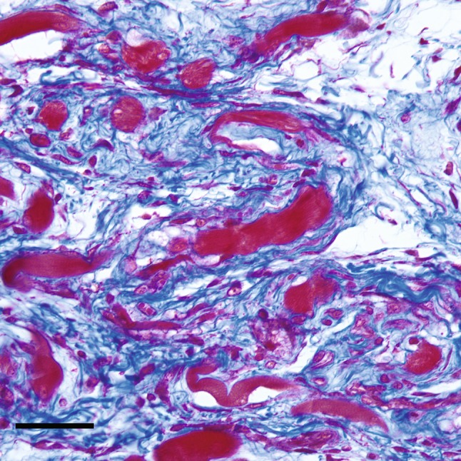
A high-magnification photomicrograph at 10 days after the induction procedure shows loose fibroconnective tissue (collagen fibers stain blue) is admixed with atrophic skeletal muscle fibers (red) (scale bar = 50 μm) (Stain, Masson’s trichrome; original magnification, ×40).