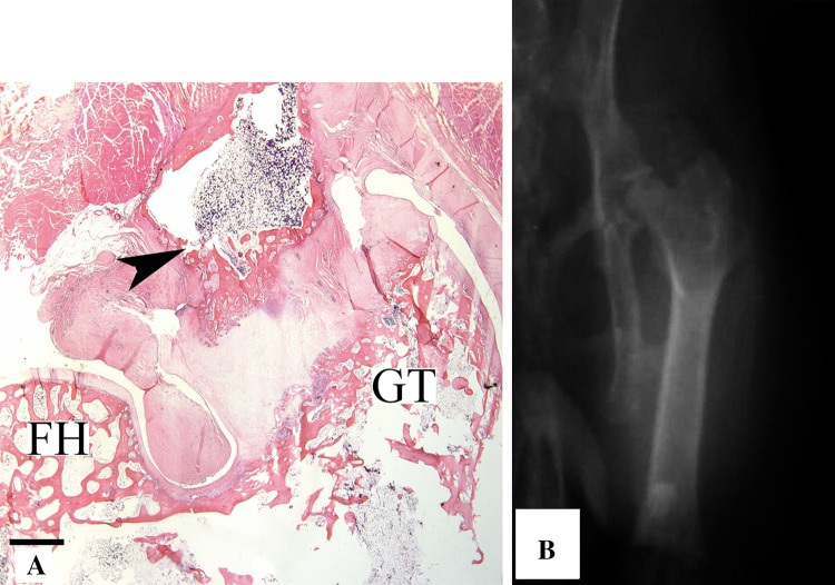
Fig. 6A–B.
(A) A low-magnification photomicrograph shows skeletal muscle 12 weeks after the induction procedure. The orientation provides observation of the head of the femur (FH) and greater trochanter (GT). A mass of heterotopic bone (arrowhead) contiguous with the femur, including marrow elements arising from hypertrophic chondrocytes in a subperiosteal location can be seen (scale bar = 1 mm) (Stain, hematoxylin and eosin; original magnification, ×1). (B) An AP radiograph of the left hip in a rabbit obtained 6 months after surgery shows HO around the hip in characteristic location.

