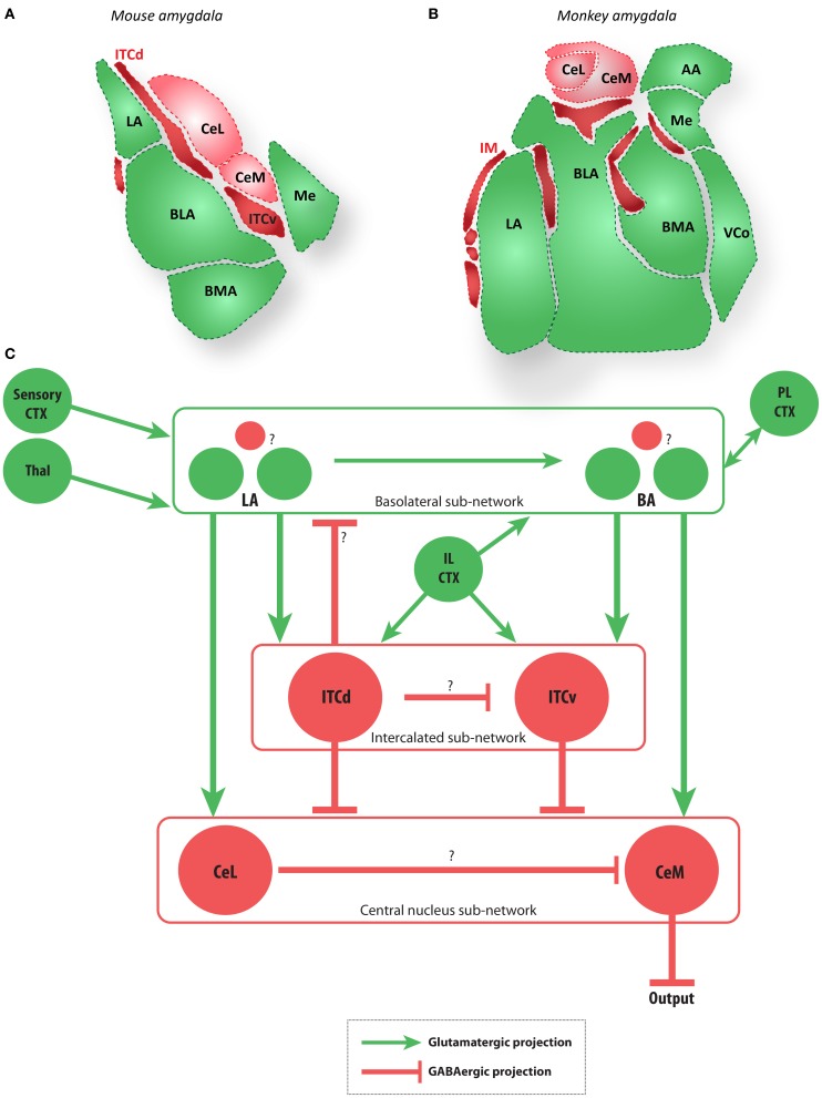
Figure 2.
The amygdala and main extrinsic and intrinsic circuits (A,B). Diagrams of coronal sections show key amygdalar nuclei in the mouse (A) and macaque monkey (B). Dark red regions are the GABAergic intercalated cells modeled here (ITCd and ITCv in mouse, and IM in macaque), light red regions are the GABAergic CeL and CeM, and green regions are glutamatergic nuclei. (C) Schematic depiction of the amygdalar local circuit. The system consists of three components: (1) the cortex-like glutamatergic basolateral sub-network (LA and BA), (2) the striatum-like GABAergic central nucleus sub-network (CeL and CeM), and (3) the GABAergic intercalated sub-network (ITCd and ITCv). The basolateral sub-network receives information about CS and US via projections from sensory cortices (CTX) and thalamus (Thal). In rodents, cortical regions IL and PL project to the intercalated and basolateral sub-networks, respectively. The CeM is a key output node of the network. Question marks indicate local microcircuit details that remain to be fully characterized experimentally. Red circles and lines represent inhibitory cell groups and connections, and green circles and lines represent excitatory cell groups and connections.

