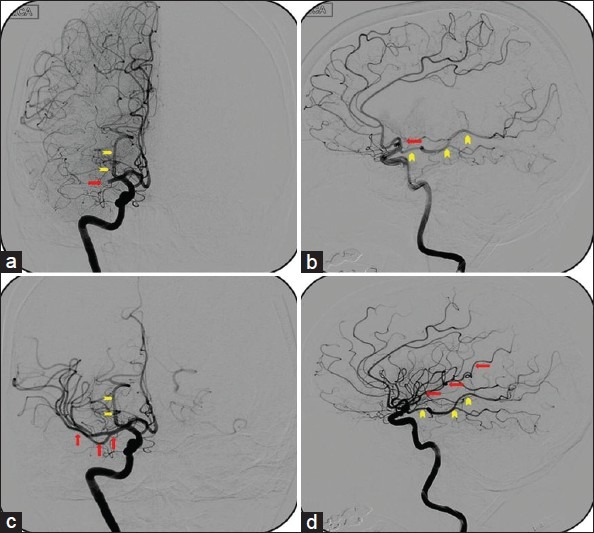
Figure 2.

Antero-posterior (a) and lateral (b) cerebral angiogram of a 60-year-old female who presented to the emergency department with symptoms of large vessel occlusion. CT scan of the head was negative for hemorrhage. CT angiogram demonstrated acute cut off of distal right M1 segment - ‘TICI grade 0’ (bold arrow). The vascular territory of fetal PCA (arrow heads) should not be confused with the MCA territory. Solitaire mechanical thrombectomy device was used to retrieve the clot form of the distal M1 segment. Post-procedure antero-posterior (c) and lateral (d) cerebral angiogram illustrating complete opacification of the distal MCA vasculature (arrows). A total of 2 passes were required to clear the clot burden and facilitate flow distally-‘TICI grade 3’
