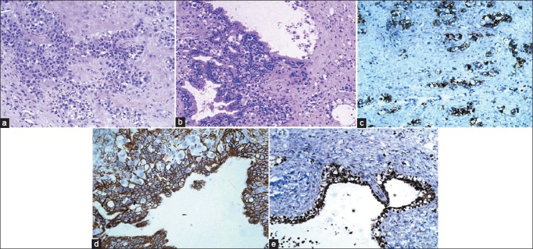
Figure 1.

(a) Suprasellar MMGCT, ×40; showing germinoma component. Cells with high N C ratio with defined cell borders and sprinkling of lymphocytes; (b) Suprasellar MMGCT, ×40; showing germinoma component admixed with glandular structures of yolk sac origin; (c) Suprasellar MMGCT, ×40; showing CD-117 positivity in germinoma component; (d) Suprasellar MMGCT, ×40; showing CK-PAN positivity in glandular structures of yolk sac origin. Note the negative germinoma cells in between; (e) Suprasellar MMGCT, ×40; showing AFP positivity in yolk sac component
