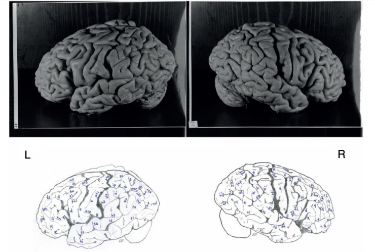
Figure 3.
Top: Photographs of the left (L) and right (R) lateral surfaces of Einstein’s brain taken from a traditional view, which lack original labels. Bottom: Our identifications. Numbers 1–4 on the right hemisphere indicate four gyri in Einstein’s right frontal lobe, rather than three as is typical. Sulci: a = additional inferior frontal; a1 = ascending branch of the superior temporal sulcus; a2 = angular; a3 = anterior occipital; aS = posterior ascending limb of the Sylvian; c = central; d = diagonal; dt = descending terminal branch of the Sylvian; e = processus acuminis; fi = inferior frontal; fm = midfrontal; fs = superior frontal; ht = posterior terminal horizontal branch of the Sylvian; inp = intermediate posterior parietal; ip = intraparietal; mf = medial frontal; ocl = lateral occipital; ocs = superior occipital; otr = transverse occipital; par = paroccipital; pci = precentral inferior; pcs = precentral superior; ps = superior parietal; pti = postcentral inferior; pts = postcentral superior; R = ascending ramus of anterior Sylvian fissure; R’ = horizontal ramus of anterior Sylvian fissure; S = Sylvian fissure; sa = sulcus acousticus; sca = subcentral anterior; scp = subcentral posterior; sip = intermedius primus of Jensen; ti = inferior temporal; tri = triangular; ts = superior temporal; tt = transverse temporal; u = unnamed. 1 = superior frontal gyrus; 2 = atypical superior middle frontal gyrus; 3 = atypical inferior middle frontal gyrus; 4 = inferior frontal gyrus (usually the ‘inferior third frontal gyrus’). K = ‘knob’ representing motor cortex for left hand. The figure is reproduced with permission from the National Museum of Health and Medicine.

