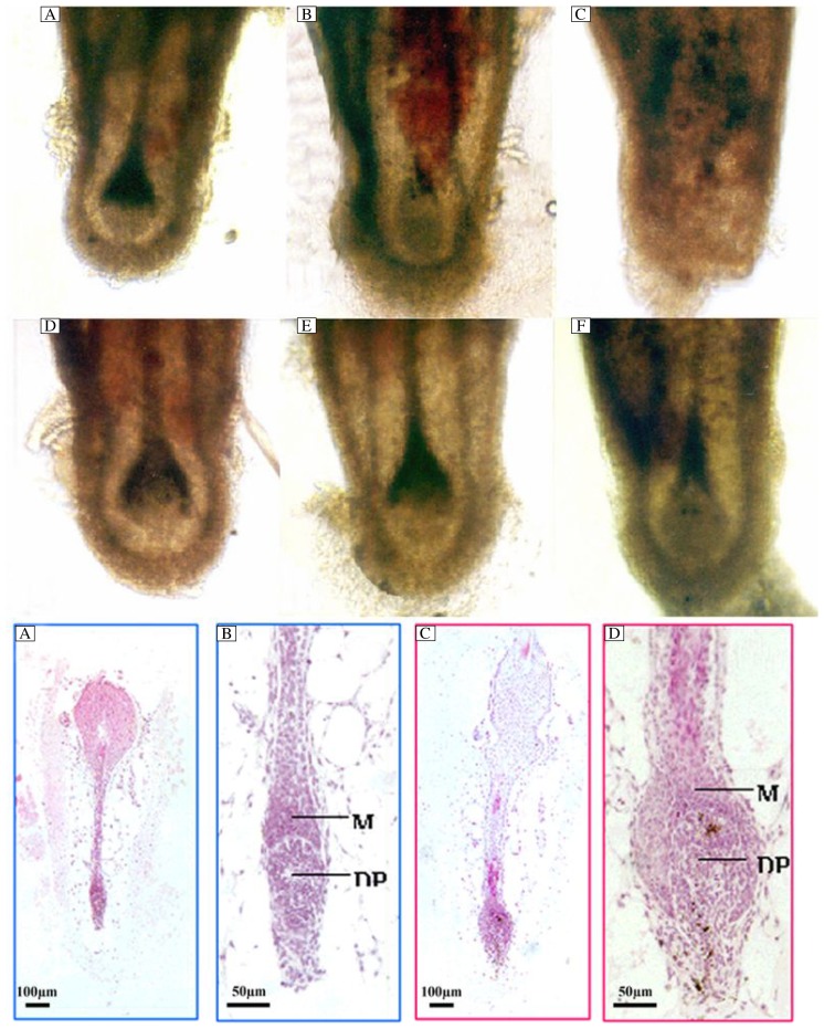
Fig. 2. Bulbs of isolated mouse vibrissae follicles on d 2 (A, D), 6 (B, E) and 10 (C, F) of culture.
Follicles are cultured with 10−7 mol/L cyclosporine A (D-F), or with vehicle (A-C). On d 10 of culture the whole shaft is either rejected from the follicle (C) or displays morphological signs in early catagen (F). Hematoxylin and eosin staining of isolated vibrissae follicles cultured for 7 d with 10−7 mol/L cyclosporine A (I, J) or vehicle (G, H). In most of vibrissae follicles treated with vehicle, the hair bulb becomes narrow and the dermal papilla is not surrounded by the epidermal matrix cells. The hair bulb looks like club hair. The dermal papilla has been compressed (G, H), while most of the vibrissae follicles treated with 10−7 mol/L cyclosporine A display full and round hair bulbs. The hair bulb is surrounded by epidermal matrix cells (I, J).

